Renal column
Extension of the cortex into the medulla
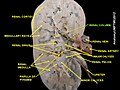
- 1. Renal pyramid
- 2. Interlobular artery
- 3. Renal artery
- 4. Renal vein
- 5. Renal hilum
- 6. Renal pelvis
- 7. Ureter
- 8. Minor calyx
- 9. Renal capsule
- 10. Inferior renal capsule
- 11. Superior renal capsule
- 12. Interlobar vein
- 13. Nephron
- 14. Renal sinus
- 15. Major calyx
- 16. Renal papilla
- 17. Renal column
[edit on Wikidata]
The renal columns, Bertin columns, or columns of Bertin, a.k.a. columns of Bertini are extensions of the renal cortex in between the renal pyramids. They allow the cortex to be better anchored. (Cortical extensions into the medullary space.)
Each column consists of lines of blood vessels and urinary tubes and a fibrous material.
A hypertrophied renal column (or renal pseudotumor) may be differentiated from an actual renal tumor with the help of a DMSA scan. The scan will show the area as one with normal activity if it is a pseudotumor or will show decreased uptake if it is a cystic or solid renal mass.
See also
Additional images
-
 Renal column
 -
 Renal column


- v
- t
- e
Anatomy of the urinary system
| Layers | |||||||
|---|---|---|---|---|---|---|---|
| Circulation | |||||||
| Nephron |
|
- Circulation
- Vesical arteries
- Vesical veins
- Vaginal artery (female)
- Detrusor muscle
- Median umbilical ligament
- Trigone
| Authority control databases |
|
|---|
 | This article related to the genitourinary system is a stub. You can help Wikipedia by expanding it. |
- v
- t
- e


